Home
/ Bone Cross Section Slide Labeled : Analysis Of New Bone Cartilage And Fibrosis Tissue In Healing Murine Allografts Using Whole Slide Imaging And A New Automated Histomorphometric Algorithm Bone Research - Each of these cylinders is called an osteon or.
Bone Cross Section Slide Labeled : Analysis Of New Bone Cartilage And Fibrosis Tissue In Healing Murine Allografts Using Whole Slide Imaging And A New Automated Histomorphometric Algorithm Bone Research - Each of these cylinders is called an osteon or.
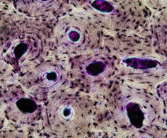
Bone Cross Section Slide Labeled : Analysis Of New Bone Cartilage And Fibrosis Tissue In Healing Murine Allografts Using Whole Slide Imaging And A New Automated Histomorphometric Algorithm Bone Research - Each of these cylinders is called an osteon or.. The basic unit of structure in compact bone is the osteon. Browse 4,294 bone cross section stock photos and images available, or search for human bone cross section to find more great stock photos and pictures. Bone cross section slide labeled : Before placing your slide on the microscope stage, remember to read the label, examine the slide with your eye and note any visible macroscopic features that might help your examination. Note that the bone matrix is deposited in concentric layers called lamellae.
There are two slides to examine. Compact bone is the denser, stronger of the two types of bone tissue ( (figure) ). Looking at a bone in cross section, there are several distinct layered regions that make up a bone. Histology spinal cord and ganglion : Bone cross section slide labeled :
Bone Tissue And Cells Under The Microscope from www.microscopemaster.com Each of these cylinders is called an osteon or. In the center of each osteon is the central canal, a space that houses blood vessels and nerves that supply bone. At this level of magnification, the fundamental structure of compact bone is visible. The periosteum contains many strong collagen fibers that are used to firmly anchor tendons and muscles to the bone for movement. Smooth muscle and endothelium in a muscular artery wall, (magnification x100). Compact bone is the denser, stronger of the two types of bone tissue ( (figure) ). This slide contained a cross section of a very small bone, and you are looking at the entire thickness of the shaft of the bone. Looking at a bone in cross section, there are several distinct layered regions that make up a bone.
Same bone section (slide 7) at higher magnification.
Fetal leg, cross section, h&e, 40x (bone marrow in tibia and fibula, developing blood cells, sinusoid 37829 x 41067, megakaryocyte 37861 x 39647, 38143 x 39087, 39555 x 36969, 31707 x 18214). Using clear epox glue, bind the section to the microscope glass slide. The central haversian canal, and horizontal canals (perforating/ volkmann's) canals contain blood vessels and nerves from the periosteum. The diaphysis and the epiphysis. That's why the color looks different. Bone matrix and cells bone matrix osseous tissue is a connective tissue and like all connective tissues contains relatively few cells and large amounts of extracellular matrix. In each osteon, the lamellae are arranged around a central haversian canal that houses nerves and blood vessels in living bone. Related posts of bone cross section labeled compact bone model labeled. In the center of each osteon is the central canal, a space that houses blood vessels and nerves that supply bone. There are two slides to examine. This is a cross section through. Looking at a bone in cross section, there are several distinct layered regions that make up a bone. Intervertebral disc, h&e, 40x (bone marrow in spongy bone of vertebrae) virtual slide.
A long bone has two parts: Very inneficient way to merge verticles. In each osteon, the lamellae are arranged around a central haversian canal that houses nerves and blood vessels in living bone. We have added a dotted line around the outside of the osteon in case you had trouble picking them out on the previous image. Smartdraw includes 1000s of professional healthcare and anatomy chart templates that you can modify and make your own.
Description from www.dartmouth.edu Notice the layered effect in the matrix. The basic unit of structure in compact bone is the osteon. Looking at a bone in cross section, there are several distinct layered regions that make up a bone. The diaphysis is the tubular shaft that runs between the proximal and distal ends of the bone. The two remodeling sites are in a bone formation phase with osteoid seams (solid arrow) and osteoblasts (open arrows) clearly visible. Note that the bone matrix is deposited in concentric layers called lamellae. Virtual slide list for histology course. Intervertebral disc, h&e, 40x (bone marrow in spongy bone of vertebrae) virtual slide.
Obtain a demineralized compact bone preparation (in cross section), preferably from the diaphysis of a long bone, and prepare to examine it microscopically.
This slide contained a cross section of a very small bone, and you are looking at the entire thickness of the shaft of the bone. Clamp the section in a vise and carefully cut it to obtain a narrow slice. The diaphysis and the epiphysis. In three dimensions an osteon is cylindrical in shape. There are two slides to examine. Note that the bone matrix is deposited in concentric layers called lamellae. Fetal leg, cross section, h&e, 40x (bone marrow in tibia and fibula, developing blood cells, sinusoid 37829 x 41067, megakaryocyte 37861 x 39647, 38143 x 39087, 39555 x 36969, 31707 x 18214). The outside of a bone is covered in a thin layer of dense irregular connective tissue called the periosteum. In each osteon, the lamellae are arranged around a central haversian canal that houses nerves and blood vessels in living bone. Virtual slide list for histology course. Each of these cylinders is called an osteon or. Consistency in fascicular organization of tibial nerve. This is a cross section through.
In this image the bar indicates the location of decalcified compact bone. Same bone section (slide 7) at higher magnification. Looking at a bone in cross section, there are several distinct layered regions that make up a bone. Consistency in fascicular organization of tibial nerve. Obtain a demineralized compact bone preparation (in cross section), preferably from the diaphysis of a long bone, and prepare to examine it microscopically.
Alveolar Bone from image.slidesharecdn.com Note that the bone matrix is deposited in concentric layers called lamellae. Notice the layered effect in the matrix. To the left is muscle tissue, and to the right is bone marrow. This photo shows a cross section through bone. The outlined area is a cross section of an osteon of compact bone. A long bone has two parts: Compact bone is the denser, stronger of the two types of bone tissue ( (figure) ). Same bone section (slide 7) at higher magnification.
In the center of each osteon is the central canal, a space that houses blood vessels and nerves that supply bone.
This slide contained a cross section of a very small bone, and you are looking at the entire thickness of the shaft of the bone. In this image the bar indicates the location of decalcified compact bone. Same bone section (slide 7) at higher magnification. Smooth muscle and endothelium in a muscular artery wall, (magnification x100). Fetal leg, cross section, h&e, 40x (bone marrow in tibia and fibula, developing blood cells, sinusoid 37829 x 41067, megakaryocyte 37861 x 39647, 38143 x 39087, 39555 x 36969, 31707 x 18214). This slide contains a section of dried compact bone. Before placing your slide on the microscope stage, remember to read the label, examine the slide with your eye and note any visible macroscopic features that might help your examination. It can be found under the periosteum and in the diaphyses of long bones, where it provides support and protection. Consistency in fascicular organization of tibial nerve. Compact bone is the denser, stronger of the two types of bone tissue ( (figure) ). Notice the layered effect in the matrix. Related posts of bone cross section labeled compact bone model labeled. This is a cross section through.
Fetal leg, cross section, h&e, 40x (bone marrow in tibia and fibula, developing blood cells, sinusoids, megakaryocytes) bone cross section. 400x this image is from a different slide than the other two images on this page.